Clinical examination tells part of the story. The rest is often only visible through imaging and having that imaging available on-site, without a referral or an extra trip across the city, changes how quickly a diagnosis can be reached and treatment can begin.
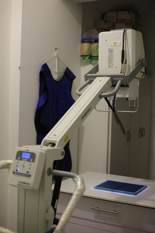
Digital X-ray
Healthy Tails operates a radiology unit at both branches. Digital X-ray is used for skeletal assessments — fractures, joint evaluations, spinal problems — as well as chest imaging to look at heart size, lung fields, and pleural changes, and abdominal views to identify foreign bodies, bladder stones, or organ abnormalities. Digital radiography produces clearer images than older film-based systems and can be reviewed and stored immediately.
Ultrasound adds a real-time, soft-tissue dimension that X-ray cannot provide. Abdominal ultrasound is used to examine the liver, gallbladder, spleen, kidneys, adrenal glands, bladder, and reproductive organs. It’s particularly valuable for evaluating masses, fluid accumulations, or organ architecture when something is off in the bloodwork but not yet obvious on physical exam.
Echocardiography (Cardiac Ultrasound)
Echocardiography — cardiac ultrasound — is available for dogs and cats presenting with heart murmurs, suspected cardiomyopathy, or symptoms that suggest a cardiac cause. It allows the team to assess chamber size, wall motion, valve function, and overall cardiac output. For breeds with known predispositions to heart disease, such as Cavalier King Charles Spaniels or Maine Coons, it serves as a critical monitoring tool.
Pre-Surgical Imaging
Pre-surgical imaging is also routine for complex cases, ensuring the team understands exactly what they’re working with before the animal goes under anesthesia. All imaging findings are interpreted in context with the full clinical picture — not read in isolation.